


1. Introduction: The Flaw May Be in the Examination
For more than a century, dystonia has been classified according to the body part in which symptoms become most visible. If the neck is affected, the disorder is called cervical dystonia. If the eyelids are affected, it is called blepharospasm. If the hand is affected during writing, it is called writer’s cramp. If symptoms emerge during instrumental performance, the diagnosis may be musician’s dystonia.
This classification is practical, familiar, and clinically useful. It allows clinicians to describe the dominant presentation, communicate clearly, and select region-specific interventions. However, usefulness should not be confused with biological completeness. The anatomical region used to classify dystonia may identify the site of greatest symptom expression rather than the full geographic extent of the underlying disorder.
In current clinical practice, the diagnosis often determines the examination, and the examination may then reinforce the diagnosis. A patient diagnosed with cervical dystonia is typically examined primarily through the neck. A musician with hand dystonia is assessed mainly through the instrument-specific hand task. A patient with blepharospasm is evaluated largely through eyelid closure, blinking, and ocular symptoms.
This approach is understandable. It focuses attention on the most disabling and visible problem. Yet it may also create a diagnostic blind spot. If the examination remains confined to the symptomatic region, subtle abnormalities in other motor systems may remain unrecognized.
When patients are examined beyond the boundaries of their clinical label, a broader set of findings may sometimes emerge. These may include altered breathing mechanics, postural asymmetries, visual tracking abnormalities, unrecognized jaw tension, abnormal eye-head coupling, changes in gait dynamics, or excessive recruitment of distant muscles during focal tasks. Traditionally, such findings may be interpreted as secondary, compensatory, incidental, or unrelated.
This article explores another possibility: that at least some of these findings may be co-manifestations of the same underlying distributed process. From this perspective, focal dystonia may not necessarily describe the biological scope of the disease. It may describe the narrowness of the standard examination.
The central question, therefore, is not whether focal dystonia exists as a useful clinical category. It clearly does. The question is whether focality reflects the true biological boundary of the disorder, or whether it identifies the site where a broader motor-network disturbance becomes most visible.
2. The Core Hypothesis
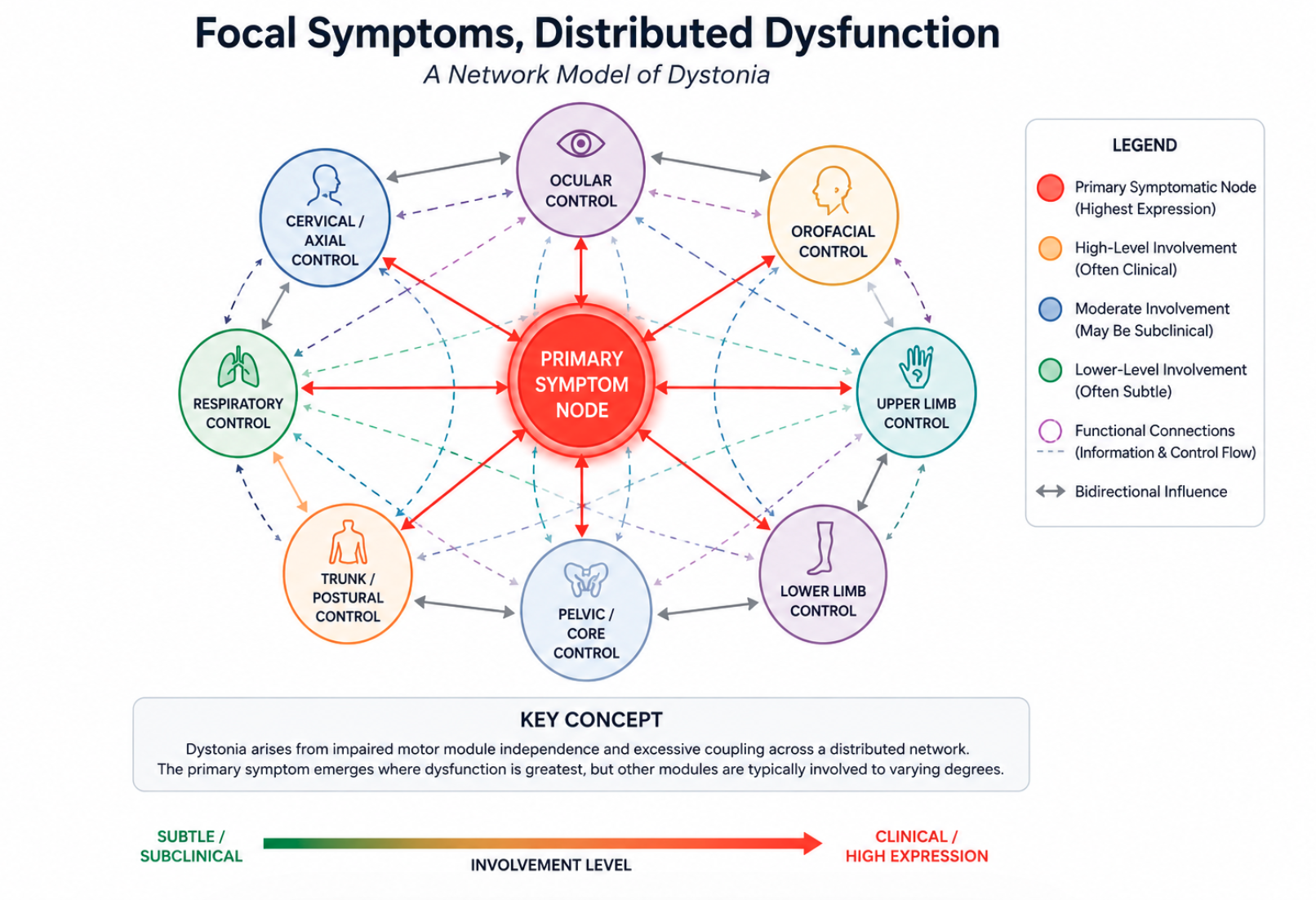
This article proposes that dystonia may be better understood, at least in many cases, as a disturbance of higher-order motor-network modulation rather than as a collection of fully separate disorders affecting isolated anatomical structures.
Under this hypothesis, cervical dystonia, blepharospasm, oromandibular dystonia, spasmodic dysphonia, musician’s dystonia, writer’s cramp, embouchure dystonia, and lower-limb dystonias would not be viewed as entirely unrelated diseases. Instead, they may represent different phenotypic expressions of a shared organizational problem. What distinguishes them may be less the fundamental nature of the neural instability and more the distribution, intensity, and task-specific expression of that instability across functional motor networks.
The visible symptom defines the clinical presentation. It may not define the full disease process.
This hypothesis does not deny the importance of local anatomy, local injury, task specificity, genetics, sensory input, peripheral feedback, or individual history. On the contrary, it proposes that these factors may help determine where a broader vulnerability becomes clinically expressed. A local trigger, repeated task demand, sensory disturbance, or biomechanical burden may lower the threshold for symptom expression in a specific region.
In this model, the clinically affected body region is understood as the point where distributed instability exceeds the threshold of visible breakdown. Other regions may remain below that threshold. They may appear normal during routine examination, yet show subtle abnormalities when specifically challenged.
This distinction is important. A disorder may be clinically focal without being biologically confined. The hand, neck, eyelids, jaw, voice, or foot may be the loudest expression of the problem, but not necessarily the only motor system involved.
3. Motor Module Independence and Pathological Coupling
Healthy movement depends on the nervous system’s ability to selectively couple and decouple different motor systems according to task demands. This capacity allows a person to move the eyes without moving the head, move the jaw without increasing neck tone, move the tongue without unnecessary jaw activation, breathe without excessive recruitment of cervical musculature, and walk without reinforcing abnormal cranial or axial posturing.
Movement is not simply generated; it is organized. The nervous system must continuously determine which motor modules should cooperate and which should remain independent. This capacity creates flexibility, precision, and freedom of movement.
A central concept in this organization is motor fractionation: the ability to produce selective movement without unwanted spread into adjacent or functionally related systems. Surround inhibition is one physiological mechanism that may contribute to this process by sharpening motor commands and reducing unnecessary activation of competing or neighboring pathways.
The Motor Module Independence framework proposes that dystonia may involve a failure of this selective independence. The problem is not only excessive muscle activity. It may also be excessive, obligatory coupling between motor modules that would ordinarily remain more independent.
Under this interpretation, a patient attempting to rotate the neck may also recruit the abdomen. A patient beginning to speak may stiffen the jaw. A patient moving one finger may involuntarily recruit the hand, forearm, shoulder girdle, or trunk. A patient performing an embouchure task may show simultaneous changes in eyelid activity, cervical tone, or respiratory bracing.
These patterns can be interpreted as motor overflow, compensation, or protective guarding. The present framework does not exclude those explanations. However, it suggests that such findings may also reflect a deeper loss of modular independence across motor systems.
The defining feature, in this model, is not merely that muscles contract too strongly. It is that motor systems become linked when the task requires them to remain differentiated. Dystonia may therefore be understood as a disorder of pathological coupling: a state in which the nervous system loses access to degrees of motor freedom that should remain available.
This interpretation generates a clinically relevant prediction: if focal dystonia involves impaired module independence, then careful testing should reveal abnormal co-activation, overflow, or coupling outside the primary symptomatic region, especially during tasks that demand high precision, intentional control, or complex fractionation.
4. The Vulnerability of Intentionality: Why Symptoms May Emerge Where They Do
If dystonia is viewed as a distributed motor-network disorder, an important question follows: why does the primary symptom emerge in one specific location rather than another?
The framework proposed here suggests that symptoms may become visible first at the node placed under the greatest functional, cognitive, emotional, sensory, or task-specific demand. Symptoms may also emerge where a local sensory-motor loop has been sensitized by trauma, overuse, inflammation, altered proprioception, or repeated high-precision demand.
In other words, the disorder may become clinically visible where the nervous system is required to perform its most difficult fractionation task, or where local stability has been compromised. The primary locus may therefore represent the point of maximum network stress rather than the complete boundary of the disorder.
This principle helps explain why different dystonia phenotypes may appear so anatomically distinct while still potentially sharing a common organizational disturbance.
Musician’s Hand Dystonia: The Price of Virtuosity
The fingers of a musician require an extraordinary degree of micro-fractionation, rapid sequencing, timing precision, and selective inhibition. Skilled performance depends on the ability to activate one motor pattern while suppressing many others. The hand must maintain speed, accuracy, independence, and adaptability under intense cognitive and sensory demands.
Because this network operates near the limits of human motor capacity, it may have a narrow margin for instability. Under conditions of high-intensity training and repeated task-specific demand, even a subtle degradation in motor-network modulation could become clinically apparent first in the finger-control modules most heavily taxed by performance.
Within this framework, musician’s dystonia is not interpreted as proof that dystonia is purely focal. Rather, it may represent an example of extreme task-specific expression: the most refined motor system fails first because it demands the highest degree of differentiation.
This model predicts that musicians with task-specific hand dystonia may show subtle abnormalities outside the hand when examined under true performance conditions. These abnormalities may be small, inconsistent, or invisible at rest, but may emerge during difficult passages, high-speed sequences, or emotionally loaded performance contexts.
Writer’s Cramp: The Cognitive Grip
Writing requires sustained, highly focused isometric control combined with fine spatial precision. Unlike many forms of hand movement, writing also involves continuous cognitive translation: language, intention, spatial planning, and motor execution must be integrated in real time.
This combination of repetitive mechanical demand and sustained intentional focus may place significant pressure on corticospinal hand networks and their supporting postural systems. Within the present framework, writer’s cramp may emerge when this high-demand motor-cognitive interface exceeds the available capacity for selective motor control.
The hand becomes the dominant site of expression because it carries the visible task. However, the model predicts that writing may also recruit abnormal activity in proximal, axial, respiratory, or orofacial modules. For example, shoulder elevation, thoracic bracing, breath-holding, jaw tension, or tongue movement may appear before or during the writing task.
These findings would not necessarily prove the framework on their own. However, if they are reproducible and measurable across patients, they would support the idea that writer’s cramp involves more than isolated hand-muscle dysfunction.
Embouchure Dystonia: The Intricate Seal
Embouchure performance requires the lips, facial muscles, tongue, jaw, respiratory system, and postural axis to cooperate with extreme precision. The performer must generate a stable seal against the mouthpiece while continuously making microscopic adjustments in pressure, tone, articulation, and airflow.
This task requires a difficult balance between rigidity and adaptability. Too little stability disrupts tone production. Too much rigidity prevents fine modulation. Under the Motor Module Independence framework, embouchure dystonia may emerge when this balance fails and the facial motor system becomes pathologically coupled to adjacent cranial, cervical, or respiratory modules.
The lips and surrounding facial muscles may become the loudest expression of the disorder because they are the point of greatest task-specific demand. However, the model predicts that examination under playing conditions may reveal additional abnormalities in breathing, jaw control, eyelid activity, cervical stabilization, or axial bracing.
Blepharospasm: Overloading Protective Systems
Blepharospasm affects a motor system that is deeply tied to protection, visual regulation, reflexive defense, emotional state, and brainstem control. The eyelids are not merely voluntary motor structures; they participate in primitive protective systems that respond to irritation, threat, light, pain, dryness, and emotional arousal.
Within the present framework, a local somatic trigger such as chronic eye irritation, inflammation, dryness, or ocular injury may increase the regulatory burden placed on eyelid-control networks. If broader motor-network modulation is already vulnerable, these protective circuits may become the first site where instability becomes clinically visible.
This interpretation does not claim that ocular irritation alone causes blepharospasm. Rather, it suggests that local sensory stress may influence where an underlying susceptibility becomes expressed.
The model predicts that patients with blepharospasm may demonstrate abnormalities beyond eyelid closure itself, including altered eye-head coordination, suboccipital recruitment, respiratory entrainment, or increased axial tone during visual tracking and blinking tasks.
Oromandibular Dystonia: The Trauma-Sensitized Node
The jaw is a highly reactive motor structure involved in speech, chewing, swallowing, facial expression, emotional regulation, airway protection, and postural organization. It also receives dense sensory input through trigeminal pathways.
In some patients, oromandibular symptoms appear after local disturbances such as dental procedures, jaw trauma, altered bite mechanics, or chronic bruxism. These associations should be interpreted cautiously, since temporal association does not prove causation. However, within the present framework, such events may act as local sensitizing factors that lower the threshold for symptom expression in the jaw-control network.
The jaw may therefore become the dominant clinical site not necessarily because the disorder is confined to the jaw, but because the local sensory-motor loop has become especially vulnerable to broader instability in motor modulation.
This hypothesis predicts that systematic examination may reveal coupling between jaw movement and cervical tone, tongue control, swallowing mechanics, breathing, or midline postural bracing.
Cervical Dystonia: The Axial Stabilizer
The neck occupies a central role in the organization of movement. It links the sensory head to the locomotor body. It helps orient the eyes, stabilize the skull, organize vestibular input, maintain equilibrium, and coordinate head position with posture and gait.
Because of this integrative role, the cervical region may be particularly sensitive to disturbances in proprioception, vestibular regulation, visual orientation, and axial postural control. Physical injury, whiplash, chronic instability, or altered local sensory feedback may increase the demand placed on cervical motor networks.
Within this framework, cervical dystonia may emerge when the systems responsible for head orientation and axial stabilization lose the capacity for selective, flexible control. The neck becomes the most visible site of expression because it is a high-demand integrative node within the motor network.
This model predicts that cervical dystonia should not be evaluated only through neck posture, range of motion, tremor, and pain. It should also be examined through eye-head coordination, respiratory mechanics, gait dynamics, pelvic dissociation, and the relationship between cervical tone and whole-body movement.
Runner’s Dystonia and Lower-Limb Dystonia: The Locomotor Threshold
Running requires precise, rhythmic, repetitive coordination across the entire body. The ankle and foot must absorb force, stabilize the body, adapt to changing surfaces, and maintain timing across thousands of cycles. Small errors in dorsiflexion, plantarflexion, inversion, or eversion can significantly disrupt locomotion.
Within the present framework, lower-limb dystonia may emerge when the locomotor network can no longer maintain selective control under repetitive high-load conditions. The foot or ankle becomes the visible site of breakdown because it is a critical output node in gait, not necessarily because the disorder is confined to that region.
Local factors such as ankle injury, altered running surface, fatigue, or biomechanical stress may influence where symptoms become expressed. However, the model predicts that broader abnormalities may also be detectable, including changes in arm swing, pelvic rotation, gaze stabilization, trunk control, and contralateral coordination.
Taken together, these examples suggest a broader principle: dystonia may become visible where the demand for motor independence is greatest, where local sensory-motor integrity has been compromised, or where a system’s margin for adaptive control is narrowest. The clinical label identifies the loudest node. It may not identify the full network.
5. Dystonia as a Disorder of Network Modulation
The framework proposed in this article suggests that dystonia may be understood not simply as a disorder of muscle activation, but as a disorder of motor-network modulation. Under this interpretation, the primary deficit is not the inability to generate movement. Rather, it is a reduced capacity to regulate movement with normal selectivity, flexibility, and independence across interacting motor systems.
Modern models of dystonia increasingly implicate distributed neural circuits involving the basal ganglia, cerebellum, thalamus, cortex, brainstem, and their reciprocal connections. Although important questions remain unresolved, there is broad agreement that dystonia cannot be fully explained by dysfunction within a single anatomical structure.
The present framework extends this network perspective by proposing that the clinical manifestations of dystonia may reflect instability within systems responsible for coordinating and modulating motor modules across the body. In this view, the disorder arises not from the failure of movement itself, but from a failure of selective organization.
Several features of dystonia appear consistent with such an interpretation. Symptoms frequently fluctuate according to stress, fatigue, emotional state, sleep quality, cognitive load, environmental context, and task complexity. Many patients demonstrate striking variability from moment to moment despite relatively preserved strength and intact basic motor capacity.
These observations suggest that modulation may be at least as important as execution. The nervous system retains the ability to generate movement, yet the quality and organization of that movement become increasingly unstable under certain conditions.
Within the Motor Module Independence framework, dystonia may therefore represent a shift away from highly differentiated control toward broader, more stereotyped patterns of recruitment. As modulation deteriorates, the nervous system may increasingly rely on evolutionarily older systems optimized for stability, orientation, and global motor organization rather than precision and fractionation.
This concept does not imply that primitive pathways are pathological. Rather, it suggests that when selective control becomes difficult to maintain, broader motor strategies may become increasingly dominant. The result may be excessive co-contraction, motor overflow, abnormal posturing, and reduced access to previously available degrees of motor freedom.
Under this interpretation, dystonic movement is not random. It represents a coherent, though maladaptive, organizational strategy. The nervous system preserves movement by sacrificing selectivity.
One practical implication of this perspective is that symptom severity should not necessarily be judged solely by the magnitude of visible movement abnormalities. Equally important may be the amount of motor freedom that has been lost. Two patients may display similar postures while possessing very different capacities for selective motor control elsewhere in the network.
This framework therefore shifts attention from the question, “Which muscles are overactive?” to the broader question, “Which motor systems have lost the ability to remain appropriately independent?”
6. The Proportionality Principle
A central structural feature of this hypothesis is the concept of proportionality.
If dystonia reflects a distributed disturbance of motor-network organization, then involvement outside the primary symptomatic region would not necessarily be absent. Instead, it may exist across a spectrum of severity ranging from clinically obvious dysfunction to subtle abnormalities that become apparent only during specific tasks or under detailed examination.
Within this framework, the apparent absence of symptoms in secondary regions does not automatically imply the absence of involvement. It may simply indicate that dysfunction remains below the threshold required for routine clinical detection.
This concept can be visualized as a gradient rather than a categorical boundary.
Traditional Model

Focal Presentation
      ↓
Pathology Confined to Target Region


Proposed Network Model

Distributed Network Disturbance
      ↓
Primary Node → High-Amplitude Expression
      ↓
Secondary Systems → Low-Amplitude Expression
      ↓
Subclinical or Task-Dependent Manifestations
Under the traditional interpretation, focal dystonia and generalized dystonia are viewed as fundamentally distinct distributions of disease. Under the proportionality model, they may instead occupy different positions along a continuum of network involvement.
Generalized dystonia would represent widespread, high-amplitude dysfunction expressed across numerous motor systems simultaneously. Focal dystonia would represent the same organizational disturbance expressed unevenly, with one dominant node crossing the threshold of visibility while other regions remain comparatively quiet.
Importantly, this framework does not argue that all forms of dystonia are equivalent in severity, mechanism, or prognosis. Rather, it proposes that apparent focality may reflect the distribution and magnitude of expression rather than the absolute presence or absence of network-wide involvement.
The proportionality principle generates several predictions. First, abnormalities outside the primary symptomatic region should be more common than currently appreciated when sufficiently sensitive testing methods are used. Second, the severity of these secondary findings should generally correlate with overall disease burden. Third, successful treatment may improve measures of network organization that extend beyond the primary body region.
These predictions are empirically testable and provide one potential avenue for evaluating the validity of the framework.
7. Clinical Observations Across Presentations
The following sections describe recurring clinical observations that appear broadly consistent with the Motor Module Independence framework. These observations should not be interpreted as established diagnostic criteria, nor should they be assumed to occur in every patient.
Rather, they represent patterns that may warrant systematic investigation. Some may ultimately prove to be compensatory adaptations. Others may reflect consequences of chronic symptoms rather than contributors to them. Nevertheless, if such findings occur with sufficient frequency and reproducibility, they could support the idea that dystonia extends beyond the boundaries of its primary clinical label.
7.1 Cervical Dystonia
Standard neurological assessment typically focuses on head position, range of motion, tremor, pain, and the pattern of cervical muscle involvement. These remain clinically important measures and often guide treatment decisions.
However, broader examination sometimes reveals additional findings involving systems outside the neck itself.
Within the framework proposed here, clinicians may observe abnormalities such as altered eye-head coordination, reduced dissociation between ocular and cervical movements, excessive recruitment of accessory respiratory musculature during quiet breathing, asymmetrical postural strategies, or gait patterns that appear to reinforce cervical posturing.
The framework predicts that these findings may represent manifestations of altered network organization rather than isolated secondary consequences of neck posture alone.
7.2 Writer’s Cramp
Writer’s cramp is traditionally viewed as a focal failure of hand control during writing. Yet careful observation often suggests involvement extending beyond the intrinsic hand musculature.
During writing tasks, some individuals appear to recruit proximal systems before visible symptoms emerge in the hand itself. Shoulder elevation, thoracic bracing, altered breathing patterns, jaw tension, and facial activation may occur during the preparation phase of writing or during periods of increased cognitive demand.
Within the present framework, these findings may represent evidence of broader motor-module coupling triggered by the writing task. The visible hand dysfunction becomes the defining symptom because it interferes directly with performance, while associated abnormalities elsewhere remain clinically secondary.
The model predicts that detailed kinematic and electromyographic studies may reveal reproducible patterns of distributed recruitment preceding overt dystonic posturing.
7.3 Musician’s Hand Dystonia
Musician’s dystonia has often been considered one of the strongest arguments for true focality because symptoms may appear only during highly specific musical passages while remaining absent during other activities.
However, the present framework offers an alternative interpretation. Because the primary task demands extraordinary levels of motor differentiation, the dominant symptom may emerge at a highly localized point even if the underlying disturbance is distributed.
Under this view, secondary abnormalities would be expected to exist but remain proportionally small. They may only become visible under genuine performance conditions.
Potential examples include altered breathing rhythm during difficult passages, excessive jaw recruitment, changes in eye-hand coordination, freezing of axial movement, or increased rigidity within distant stabilizing systems.
The framework predicts that such findings should become more apparent as task complexity and performance pressure increase.
7.4 Embouchure Dystonia
Clinical attention in embouchure dystonia is understandably directed toward the lips, facial musculature, and instrument-specific performance deficits. Yet performance itself depends upon the coordinated interaction of multiple systems extending beyond the embouchure.
Within the present framework, embouchure dysfunction may represent the visible endpoint of broader abnormalities involving respiratory control, cervical stabilization, cranial motor coordination, and postural organization.
Some performers appear to demonstrate involuntary blinking, altered facial recruitment patterns, cervical stiffening, or abdominal co-contraction during difficult passages. Whether these findings represent compensatory behavior, downstream consequences, or manifestations of broader network dysfunction remains uncertain.
The framework predicts that at least a subset of these findings may prove reproducible under formal investigation.
7.5 Blepharospasm
Blepharospasm is typically defined through involuntary eyelid closure and abnormalities of blinking. Yet patients frequently report symptoms that extend beyond the eyelids themselves.
Expanded examination may reveal altered eye-head coordination, excessive recruitment of cervical musculature during visual tasks, changes in head posture, or symptom fluctuations associated with respiratory cycles and attentional state.
Within the proposed model, these observations may reflect broader disturbances within orienting networks responsible for integrating visual, vestibular, cervical, and postural control.
This interpretation predicts that future studies may identify measurable abnormalities in systems that participate in gaze stabilization and head orientation even when the primary complaint remains eyelid spasm.
7.6 Oromandibular Dystonia
Oromandibular dystonia is often described through abnormalities of jaw opening, jaw closing, tongue movement, chewing, or speech production. However, these functions exist within a densely interconnected motor network involving cranial, cervical, respiratory, and postural systems.
Patients may demonstrate changes in cervical muscle activity during jaw movement, alterations in swallowing mechanics, excessive tongue recruitment, changes in breathing patterns, or generalized midline bracing during periods of symptom amplification.
The framework predicts that these findings may represent examples of pathological coupling extending beyond the primary site of symptom expression.
7.7 Runner’s Dystonia and Lower-Limb Presentations
Lower-limb dystonias are frequently interpreted through an orthopedic or localized motor lens because symptoms appear most visibly in the foot, ankle, or leg. Yet locomotion is inherently a whole-body activity requiring coordination across multiple levels of the motor system.
Some patients appear to develop alterations in arm swing, trunk rotation, pelvic dissociation, gaze stabilization, and postural organization before the dystonic movement becomes fully apparent.
Within the present framework, these findings may indicate that locomotor dystonia reflects broader disturbances in network coordination rather than isolated dysfunction of a single limb segment.
The model predicts that future gait-analysis studies may reveal abnormalities distributed across the locomotor chain that precede or accompany the visible lower-limb symptoms.
Taken together, these observations do not establish the framework as correct. They do, however, suggest a coherent question worthy of systematic investigation: when patients diagnosed with focal dystonia are examined beyond the boundaries of the primary symptom region, how often does evidence of broader motor-network involvement emerge?
8. The Diagnostic Blind Spot: Toward an Expanded Examination
If dystonia reflects a broader disturbance of motor-network organization rather than dysfunction confined entirely to a single body region, then the scope of clinical examination may require expansion.
This proposal does not imply that traditional neurological assessment is inadequate. Current examination protocols are highly effective for identifying dystonia, characterizing phenomenology, and guiding treatment. However, they are primarily designed to identify where symptoms are most visible. They may be less effective at detecting low-amplitude manifestations distributed elsewhere in the motor system.
Within the framework proposed here, the absence of examination is not equivalent to the absence of involvement. A system that is never challenged, measured, or observed under relevant task conditions may appear normal despite contributing to the broader clinical picture.
The central diagnostic question therefore shifts from:
Which body part is affected?
to:
How extensively has motor-module independence been compromised throughout the network?
To address this question, a broader examination strategy may be required.
| Domain | Assessment Method | Potential Findings |
|---|---|---|
| Visual Motor Control | Video-oculography, smooth pursuit testing, saccadic assessment | Abnormal eye-head coupling, excessive cervical recruitment during isolated ocular tasks |
| Respiratory Motor Control | Breathing observation, inductive plethysmography, respiratory kinematics | Loss of diaphragmatic leadership, accessory muscle over-recruitment, paradoxical bracing |
| Locomotor Organization | Gait analysis, force-plate assessment, motion capture | Reduced thoracic-pelvic dissociation, gait-related amplification of dystonic patterns |
| Orofacial Function | Tongue, jaw, and swallowing assessment | Abnormal coupling between tongue, jaw, cervical, and respiratory systems |
| Motor Module Independence | Surface EMG across multiple body regions | Unexpected activation of distant muscle groups during focal tasks |
Importantly, the purpose of such assessment would not be to demonstrate that every patient possesses every abnormality. Rather, it would be to determine whether distributed involvement is more common than currently appreciated and whether identifiable patterns exist across different dystonia phenotypes.
If the framework is correct, future diagnostic approaches may increasingly focus on network-wide organization rather than solely on the dominant symptomatic region.
9. Therapeutic and Rehabilitation Implications
Redefining Neurorehabilitation
Traditional rehabilitation approaches often focus on the most visibly affected muscles or body regions. This strategy is understandable because those regions produce the symptoms that interfere most directly with function.
However, if dystonia involves impaired motor-module independence, then rehabilitation may need to target the restoration of selective control rather than simply the reduction of excessive activity.
Within this framework, the therapeutic objective is not merely relaxation of overactive muscles. The objective is the recovery of lost degrees of motor freedom.
Interventions would therefore emphasize the deliberate separation of motor systems that have become pathologically linked. Rehabilitation becomes an exercise in decoupling.
For example, a patient with cervical dystonia might practice visual tracking while maintaining relaxed cervical control. A patient with writer’s cramp might train breathing independence during writing tasks. A musician might work on restoring respiratory, ocular, and postural freedom during performance-specific movements.
The goal is not simply to suppress symptoms. The goal is to increase the nervous system’s capacity for differentiated motor behavior.
This approach generates an important prediction: successful rehabilitation should improve measures of motor independence beyond the primary symptomatic body region.
A New Interpretation of Botulinum Toxin
Botulinum toxin remains one of the most effective treatments currently available for many forms of focal dystonia. Traditionally, its therapeutic value has been understood primarily through its ability to reduce excessive muscular activity at the symptomatic site.
The present framework does not challenge this understanding. However, it offers an additional interpretation.
By reducing activity within the dominant symptomatic node, botulinum toxin may temporarily decrease the volume of aberrant sensory feedback returning to central motor networks. This may reduce the influence of the most disruptive source of network instability.
Under this interpretation, botulinum toxin functions not only as a local intervention but also as a potential facilitator of broader motor reorganization.
The resulting therapeutic window may provide an opportunity for targeted rehabilitation aimed at restoring motor-module independence throughout the network.
This hypothesis generates a testable prediction: patients who combine botulinum toxin treatment with network-oriented rehabilitation may demonstrate improvements that extend beyond the direct effects of local muscle weakening.
10. Conclusion
Dystonia has long been categorized according to where symptoms become most visible. This approach remains clinically useful and continues to provide an effective framework for diagnosis and treatment.
Yet an important question remains open.
Does the anatomical region used to define the diagnosis truly define the biological boundaries of the disorder?
The framework proposed in this article suggests that it may not.
Under the Motor Module Independence model, dystonia is interpreted as a disorder of distributed motor-network modulation characterized by reduced motor fractionation and excessive coupling between systems that would ordinarily remain more independent.
The primary symptomatic region represents the point at which dysfunction becomes most visible. It may not represent the full extent of involvement.
This interpretation offers a possible explanation for why many patients appear to exhibit abnormalities extending beyond their formal clinical classification and why symptoms often fluctuate according to context, task demand, cognitive load, and emotional state.
Most importantly, it generates a set of predictions that can be tested empirically.
If those predictions are supported, the concept of focal dystonia may require revision. If they are not supported, the framework should be modified or rejected accordingly.
The purpose of this model is therefore not to provide a final answer. Its purpose is to propose a question that has received surprisingly little systematic attention:
Is focal dystonia truly focal, or does focality reflect the limits of how we currently examine the disorder?
11. Predictions Generated by the Framework
A useful scientific framework should generate predictions that can be tested objectively. The Motor Module Independence framework produces several specific predictions.
Prediction 1: Secondary Abnormalities Will Be Common
Patients diagnosed with focal dystonia will frequently demonstrate measurable abnormalities outside the primary symptomatic region when examined with sufficiently sensitive methods.
Prediction 2: Severity Will Scale Across Systems
The severity of secondary findings will generally correlate with the severity of the dominant symptom, reflecting a common underlying disturbance rather than unrelated deficits.
Prediction 3: Different Phenotypes Will Share Common Features
Despite affecting different body regions, cervical dystonia, writer’s cramp, embouchure dystonia, blepharospasm, and lower-limb dystonias will demonstrate overlapping abnormalities in motor-module independence.
Prediction 4: Distributed Changes Will Precede Visible Symptoms
Subclinical abnormalities may be detectable before overt symptom expression, particularly in individuals at elevated risk due to genetics, intensive skill acquisition, or prior dystonic symptoms elsewhere.
Prediction 5: Successful Treatment Will Improve Network Organization
Effective interventions will improve not only the primary symptom but also broader measures of motor coordination, motor fractionation, and network independence.
Prediction 6: Task Complexity Will Amplify Hidden Dysfunction
Secondary abnormalities will become more apparent as cognitive demand, emotional stress, motor complexity, or environmental challenge increases.
Prediction 7: Motor Overflow Reflects a General Principle
Motor overflow observed in dystonia will prove to be one manifestation of a broader impairment in selective motor-network organization.
12. Potential Falsification Criteria
A scientific framework must also define the conditions under which it could be rejected.
The Motor Module Independence framework would be weakened if the following observations consistently occurred:
- Expanded examination protocols fail to identify abnormalities outside the primary symptomatic region.
- Secondary findings are fully explained by compensation, pain, biomechanical adaptation, or psychological factors without evidence of shared network involvement.
- Patients with different dystonia phenotypes do not demonstrate common abnormalities in motor-module organization.
- Surface EMG, motion capture, respiratory assessment, gait analysis, and oculomotor testing fail to reveal evidence of abnormal coupling outside the primary dystonic task.
- Network-oriented rehabilitation produces no measurable advantages compared with conventional focal approaches.
- Improvement of the primary symptom fails to produce any detectable changes in broader measures of motor organization.
If these outcomes are consistently demonstrated, the framework would require substantial revision or abandonment.
Conversely, if future studies repeatedly identify distributed abnormalities across multiple dystonia phenotypes and show that treatment influences network-wide measures of motor organization, support for the model would increase.
The value of the framework therefore rests not in its elegance but in its ability to generate predictions that can be rigorously tested.